|
| |
| |
|
Problems
that occur with a woman’s reproductive organs
sometimes cannot be found by a physical examination
alone. Laboratory tests, Ultrasound, X- Rays may still
leave some uncertainty. Frequently, problems that cannot
be discovered by routine investigations can be discovered
by laparoscopy or hysteroscopy, two procedures which
provide a direct look at the pelvic organs. Laparoscopy
and hysteroscopy can be used for both diagnostic (looking
only) and operative (looking and treating) purposes.
Diagnostic laparoscopy may be recommended to look at
the outside of the uterus, fallopian tubes, ovaries,
and internal pelvic area. Diagnostic hysteroscopy is
used to look inside the uterus. If an abnormal condition
is detected during the diagnostic procedure, operative
laparoscopy or hysteroscopy can often be performed to
correct it at the same time, avoiding the need for second
surgery.
DIAGNOSTIC LAPAROSCOPY
Laparoscopy can help gynecologists diagnose many problems
including endometriosis, uterine fibroids and other
structural abnormalities, ovarian cysts, adhesions,
ectopic pregnancy, tubal disease, and genital tuberculosis.
Many infertile patients require laparoscopy for a complete
evaluation. Generally, the procedure is performed after
the basic infertility tests, although the presence of
pain, history of past infection or an abnormal ultrasound
may signal a need to perform diagnostic laparoscopy
sooner in the evaluation.
Laparoscopy is usually performed as an outpatient basis,
under general anesthesia, and with minimal discomfort.
LAPAROSCOPIC
PROCEDURE
After anesthesia, a needle is inserted through the navel,
and the abdomen is filled with carbon dioxide gas. As
the gas enters the abdomen, it creates a space inside
by pushing the abdominal wall and the bowel away from
the organs in the pelvic area allowing a view of the
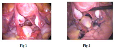
reproductive organs. Next, a long thin telescope (laparoscope)
is inserted through the insertion in the navel. It is
connected to a tiny camera which sends images to a television
monitor. While looking at the monitor, the surgeon can
see the uterus, fallopian tubes, ovaries, and nearby
structures (figure 1). A small probe is inserted through
another incision in order to move the pelvic organs
into clear view Additionally, a blue solution is injected
through the cervix to determine if the fallopian tubes
are open. (fig 2). If no abnormalities are noted at
this time, one or two stitches close the incisions.
The incisions are closed using an adhesive dressing.
If defects or abnormalities are discovered, one can
proceed to operative laparoscopy.
 OPERATIVE LAPAROSCOPY
Many infertility disorders can be safely treated through
the laparoscope at the same sitting. Operating instruments
like graspers, biopsy forceps, scissors, coagulators,
electrosurgical or laser instruments, needle holders
and suture materials are inserted through two or three
incisions in the area above the pubis.
Operative procedures include adhesiolysis, treatment
of blocked tubes, fulguration of endometriosis, removal
of chocolate cysts, treatment of ovarian cysts, PCOD
drilling, removal of diseased ovaries, removal of uterine
fibroids, and treatment of ectopic pregnancy.
Operations for female sterilization, hysterectomy, urinary
incontinence and genital prolapse can also be performed
laparoscopically.
|
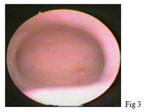
DIAGNOSTIC HYSTEROSCOPY
Hysteroscopy is an important tool in the study of infertility,
recurrent miscarriage, or abnormal uterine bleeding.
Diagnostic hysteroscopy is used to examine the inside
of the uterus, also known as the uterine cavity (figure
3) and is helpful in diagnosing abnormal uterine conditions
such as polyps, internal fibroids, scarring, and developmental
abnormalities. A hysterosalpingogram (an x-ray of the
uterus and fallopian tubes) may be performed before
a diagnostic hysteroscopy. Diagnostic hysteroscopy is
usually conducted on an outpatient basis with either
general or local anesthesia.
For infertility evaluation the hysteroscopy
and laparoscopy are combined together usually soon after
menstruation because the uterine cavity is more easily
evaluated and there is no risk of interrupting a pregnancy.
|
|
HYSTEROSCOPIC PROCEDURE
After dilating the cervix (mouth of the uterus) with
a series of dilators, a narrow telescope (hysteroscope)
is passed through the cervix into the uterine cavity.
Special clear solutions are then injected into the uterus
through the hysteroscope sheath. This distends the uterine
cavity, clears blood and mucus, and allows the gynecologist
to directly view the internal structure of the uterus.
OPERATIVE HYSTEROSCOPY
A wider hysteroscope allow operating instruments such
as scissors, biopsy forceps, graspers, electrosurgical
or laser instruments to be introduced into the uterine
cavity through a channel in the operative hysteroscope.
Fibroids, polyps, adhesions can be removed from inside
the uterus. Congenital abnormalities, such as uterine
septum, can also be corrected through the hysteroscope.
After surgical repair, a Foley catheter or intrauterine
device may be placed inside the uterus to prevent the
uterine walls from fusing together. Antibiotics and/or
hormonal medication may also be prescribed after uterine
surgery to prevent infection and stimulate healing of
the endometrium (uterine lining).
RISKS OF LAPAROSCOPY / HYSTEROSCOPY
Serious complications of diagnostic and operative laparoscopy
are rare. Allergic reactions and anesthesia complications
rarely occur. The major risk is damage to the bowel,
bladder, ureters, major blood vessels, or other organs,
which would require immediate laparotomy to repair the
injury. Injury can also occur during the insertion of
various instruments through the abdominal wall or during
operative treatment. Certain conditions may increase
the risk of serious complications. These include previous
abdominal surgery, presence of bowel or pelvic adhesions,
severe endometriosis, obesity or excessive thinness.
In experienced hands the risk of injury is 2-3 per 1000
procedures.
The risk of death during laparoscopy is 1-2 per 100000
procedures, is less than the risk of death during pregnancy.
Complications of hysteroscopy are rare and seldom serious.
Perforation of the uterus (hole in the uterus) is the
most common complication, but the hole usually heals
on its own. Some complications related to the liquids
used to distend the uterus include fluid overload, pulmonary
edema (fluid in the lungs), blood clotting problems,
and severe allergic reactions. Complications related
to the surgical procedure include damage to intra-abdominal
organs and hemorrhage. Severe or life threatening complications,
however, are very uncommon.
RECOVERY
After Laparoscopy / Hysteroscopy, the patient is allowed
to rest for 2 – 4 hours to recover from the anesthesia.
She is allowed liquids after 4 hours and soft diet in
the evening. After the operation, the patient may feel
some discomfort :
-
Mild nausea from medication /
anesthesia
-
A sore throat if a breathing
tube was used during anesthesia
-
Pain in the shoulders from the
gas used during laparoscopy
-
Pain at the site of incisions
-
Cramps, like menstrual cramps
-
Discharge like menstrual flow
for a few days
Most of these minor complaints are
gone in a day or two after surgery.
CONCLUSION
Previously, diagnosing and treating gynecological problems
required major surgery and many days of hospitalization.
However, laparoscopy and hysteroscopy allows correction
of these problems on an outpatient basis. The procedures
decrease patient discomfort, significantly reduce recovery
time and has minimal risks. |
|
| |
|
|